Clubfoot
What is clubfoot?
Congenital Talipes Equinovarus (CTEV), often referred to as clubfoot, is a congenital deformity where one or both feet are twisted inward and downward (Fig.1), giving the foot a characteristic inward and backward appearance.
This condition can vary in severity, potentially affecting the muscles, bones, and joints of the foot, and may occur in one or both feet.
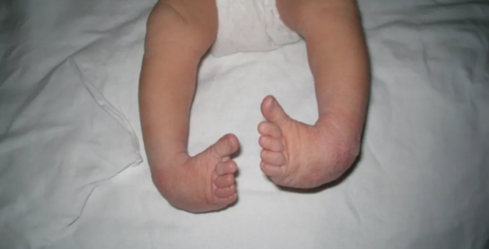
Fig 1. Congenital clubfoot
There are two types of CTEV:
Postural CTEV: Primarily due to muscle imbalance and tightness. It is usually mild and does not involve structural abnormalities of the bones or joints.
Structural CTEV: A more severe form that involves the bones and joints of the foot. In this type, the foot’s position is rigid and cannot be easily adjusted or moved into a normal range of motion, even with external force.
Causes of clubfoot
The exact cause of clubfoot is not entirely understood but may result from a combination of genetic, environmental, and mechanical factors.
Signs and symptoms of clubfoot
The condition is often quite obvious after the birth of your child. The physical appearance of the foot may vary.
Signs and symptoms include:
- One or both feet turning inwards
- Tightness in calf muscles
- The calf muscle and affected foot may be slightly smaller than normal
- Decreased joint range of movement in the foot (for structural CTEV)
Treatment for clubfoot
Before initiating treatment, babies with clubfoot are typically referred to a physiotherapist or specialist who will conduct a thorough assessment to determine the type and severity of CTEV affecting your child. This evaluation helps guide the treatment approach to achieve the best possible outcome.
Here are the common treatments for the two types of clubfoot:
Postural CTEV: This type often resolves on its own with minimal intervention. However, physiotherapy may be recommended to assist with stretching exercises and stimulation techniques to encourage proper foot alignment and muscle development.
Structural CTEV: Serial casting and manipulation with minimal surgery if necessary. For the most severe deformities, comprehensive surgery may be required to correct bone and joint abnormalities.
Early and consistent treatment is essential for correcting the deformity and ensuring that the child develops normal walking and foot function.
Method of treatment
We practice the Ponseti method of treatment.
The objectives are:
- To correct the deformity early
- To correct the foot to achieve full function
- To maintain the correction until the child is fully grown
- To minimise surgery
The length of treatment may require a minimum of four years depending on the response.
Serial manipulation and casting
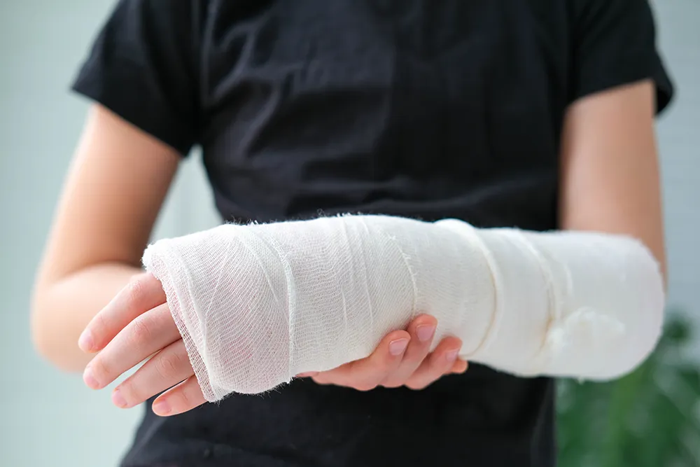
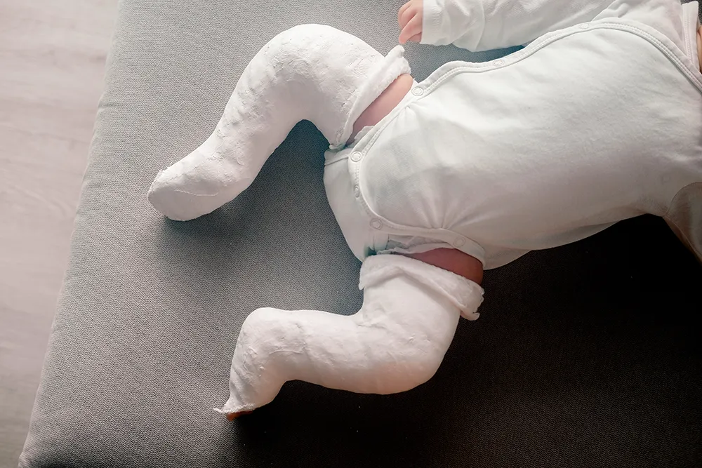
Your child’s foot will be placed in a cast from toe to groin to gradually correct the shape of the foot and try to prevent future deformity.
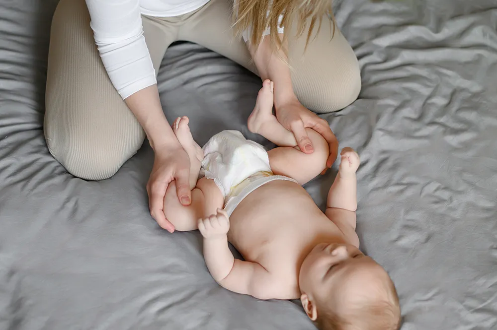
After the foot is in this position for about a week, the muscles and ligaments will stretch enough to make further corrections possible. The cast is then removed and the same process of gentle manipulation is repeated approximately five to six times until adequate correction has been achieved.
In some cases, to achieve full correction, the heel cord (Achilles tendon) is clipped (tenotomy) before the application of the last plaster cast. The tendon re-attaches with no weakness in two to three weeks.
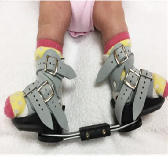
Fig 2. Special boots and bars
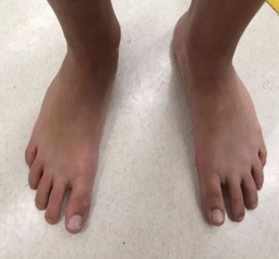
Fig 3. A fully corrected right-sided clubfoot
of a 15 years old
